Anaphase Drawing
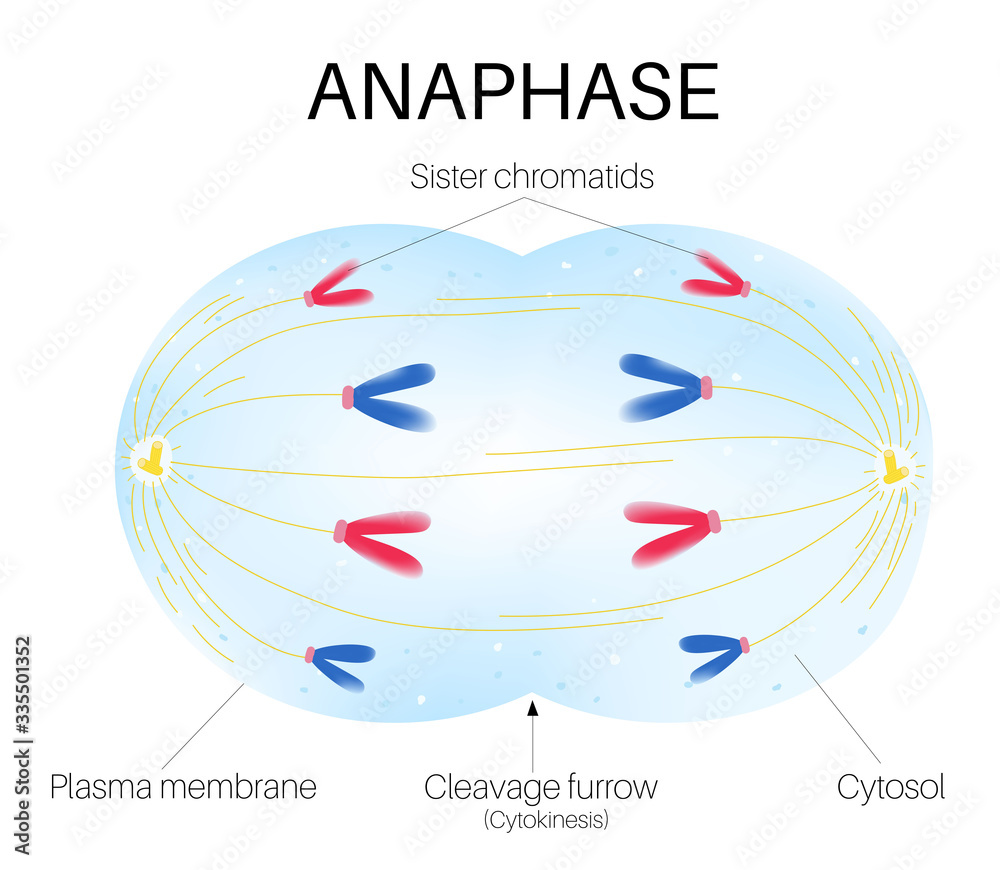
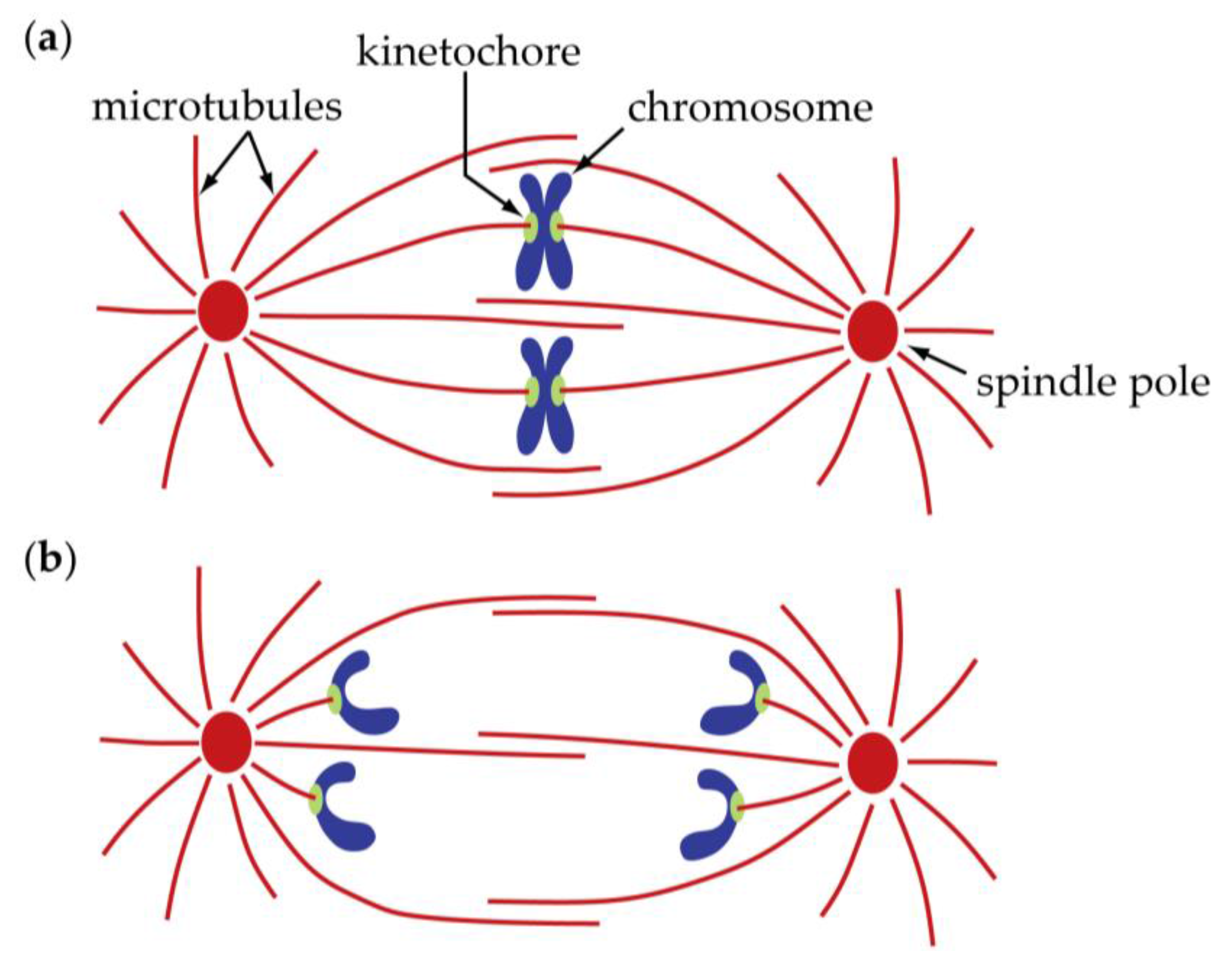
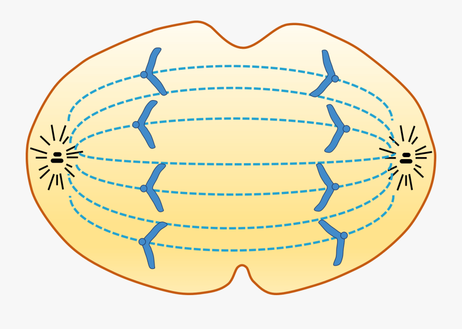
Anaphase Drawing - Web anaphase i is the third stage of meiosis i and follows prophase i and metaphase i. It starts as soon as metaphase ends, and it comes right before telophase. Each chromatid, now called a chromosome, is pulled rapidly toward the centrosome to which its microtubule was attached. Web generally, anaphase i involve separating the chromosomes from each sister chromatid to the opposite poles still attached to the microtubules of the cell while anaphase 2 involves the actual split of the sister chromatids into single chromatids. Web anaphase begins as sister chromatids separate, drawn toward each centrosome by spindle fibers of the spindle apparatus. Web model anaphase by removing the white beads (centromere) from the sister chromatids to separate and move them toward opposite poles of the cell. At the end of anaphase, each pole contains a complete compilation of chromosomes. Web during anaphase, the sister chromatids at the equatorial plane are split apart at the centromere. Prophase (sometimes divided into early prophase and prometaphase), metaphase, anaphase, and telophase. Humans have 46) but the diagrams below show mitosis of an animal cell with only four chromosomes, for simplicity. Web during anaphase, the sister chromatids at the equatorial plane are split apart at the centromere. Centrosomes and microtubules play pivotal roles in orchestrating this complex process, ensuring the successful replication of cells. The stage before anaphase, metaphase, the chromosomes are pulled to the metaphase plate, in the middle of the cell. In cytokinesis, the cytoplasm of. Web anaphase is a stage during eukaryotic cell division in which the chromosomes are segregated to opposite poles of the cell. Web mitosis, a key part of the cell cycle, involves a series of stages (prophase, metaphase, anaphase, and telophase) that facilitate cell division and genetic information transmission. Spindle fibers are green, chromosomes are blue, and kinetochores are pink. Web generally, anaphase i involve separating the chromosomes from each sister chromatid to the opposite poles still attached to the microtubules of the cell while anaphase 2 involves the actual split of the sister chromatids into single chromatids. After separation at the centromere, the chromatids are now called chromosomes. Web mitosis takes place in four stages: In metaphase i, chromosomes line up in the middle of the cell. After separation at the centromere, the chromatids are now called chromosomes. Web anaphase, in mitosis and meiosis, the stage of cell division in which separated chromatids (or homologous [like] chromosome pairs, as in the first meiotic division) move toward the opposite poles of the spindle apparatus. Centrosomes and microtubules play pivotal roles in orchestrating this complex process, ensuring the successful replication of cells. Web in anaphase, the paired chromosomes ( sister chromatids) separate and begin moving to opposite ends (poles) of the cell. Web anaphase is a stage during eukaryotic cell division in which the chromosomes are segregated to opposite poles of the cell. These phases are prophase, prometaphase, metaphase, anaphase, and telophase. You can learn more about these stages in the video on mitosis. During prophase i, chromosomes pair up and exchange genetic material, creating more variation. Web the four stages of mitosis are known as prophase, metaphase, anaphase, telophase. After separation at the centromere, the chromatids are now called chromosomes. Web anaphase is the fourth phase of mitosis, which is a process that separates the duplicated genetic material carried in the nucleus of a parent cell into two, identical daughter cells. These phases are prophase, prometaphase, metaphase, anaphase, and telophase. Prophase (sometimes divided into early prophase and prometaphase), metaphase,. After separation at the centromere, the chromatids are now called chromosomes. Web mitosis consists of five morphologically distinct phases: The process of mitosis is significant in both cell division as well as cell reproduction. Spindle fibers not connected to chromatids lengthen and elongate the cell. Web anaphase, in mitosis and meiosis, the stage of cell division in which separated chromatids. Prophase, metaphase, anaphase, and telophase. Web anaphase is the fourth phase of mitosis, which is a process that separates the duplicated genetic material carried in the nucleus of a parent cell into two, identical daughter cells. The chromatids only start separating when the pressure is sufficient to split the centromere. Web during anaphase, the sister chromatids at the equatorial plane. Web generally, anaphase i involve separating the chromosomes from each sister chromatid to the opposite poles still attached to the microtubules of the cell while anaphase 2 involves the actual split of the sister chromatids into single chromatids. This stage is characterized by the movement of chromosomes to both poles of a meiotic cell via a microtubule network known as. Web anaphase, in mitosis and meiosis, the stage of cell division in which separated chromatids (or homologous [like] chromosome pairs, as in the first meiotic division) move toward the opposite poles of the spindle apparatus. The major event of anaphase, the third phase of mitosis, is the sister chromatids moving to opposite poles of the cells due to the action. Most organisms contain many chromosomes in the nuclei of their cells (eg. The process of mitosis is significant in both cell division as well as cell reproduction. Web today, mitosis is understood to involve five phases, based on the physical state of the chromosomes and spindle. Web anaphase, in mitosis and meiosis, the stage of cell division in which separated. Web anaphase, in mitosis and meiosis, the stage of cell division in which separated chromatids (or homologous [like] chromosome pairs, as in the first meiotic division) move toward the opposite poles of the spindle apparatus. Web this is a drawing of anaphase and a real photomicrograph of a cell in anaphase. Prophase, metaphase, anaphase, and telophase. The process of mitosis. During prophase i, chromosomes pair up and exchange genetic material, creating more variation. This stage is characterized by the movement of chromosomes to both poles of a meiotic cell via a microtubule network known as the spindle apparatus. Web mitosis consists of four basic phases: Web model anaphase by removing the white beads (centromere) from the sister chromatids to separate. Spindle fibers not connected to chromatids lengthen and elongate the cell. Web anaphase begins as sister chromatids separate, drawn toward each centrosome by spindle fibers of the spindle apparatus. It starts as soon as metaphase ends, and it comes right before telophase. In metaphase i, chromosomes line up in the middle of the cell. Prophase, metaphase, anaphase, and telophase. Web today, mitosis is understood to involve five phases, based on the physical state of the chromosomes and spindle. Web mitosis consists of five morphologically distinct phases: During prophase i, chromosomes pair up and exchange genetic material, creating more variation. Web anaphase begins as sister chromatids separate, drawn toward each centrosome by spindle fibers of the spindle apparatus. Web anaphase. In metaphase i, chromosomes line up in the middle of the cell. The process of mitosis is significant in both cell division as well as cell reproduction. Web this is a drawing of anaphase and a real photomicrograph of a cell in anaphase. After separation at the centromere, the chromatids are now called chromosomes. During prophase i, chromosomes pair up and exchange genetic material, creating more variation. This stage is characterized by the movement of chromosomes to both poles of a meiotic cell via a microtubule network known as the spindle apparatus. Web mitosis consists of five morphologically distinct phases: In cytokinesis, the cytoplasm of. Web mitosis, a key part of the cell cycle, involves a series of stages (prophase, metaphase, anaphase, and telophase) that facilitate cell division and genetic information transmission. Web anaphase begins as sister chromatids separate, drawn toward each centrosome by spindle fibers of the spindle apparatus. Most organisms contain many chromosomes in the nuclei of their cells (eg. Prophase, prometaphase, metaphase, anaphase, and telophase. Prophase i, metaphase i, anaphase i, and telophase i. It starts as soon as metaphase ends, and it comes right before telophase. You can learn more about these stages in the video on mitosis. Web anaphase is the fourth phase of mitosis, which is a process that separates the duplicated genetic material carried in the nucleus of a parent cell into two, identical daughter cells.Anaphase stage of mitosis 8576777 Vector Art at Vecteezy
What Is Anaphase in Cell Biology?
Anaphase Illustrations Illustrations, RoyaltyFree Vector Graphics
Meiosis Anaphase Free Images at vector clip art online
Anaphase is the phase of the cell cycle Stock Vector Adobe Stock
9+ which diagram represents anaphase i of meiosis YenyukLilia
Biology Free FullText Anaphase A Disassembling Microtubules Move
Anaphase — Definition & Diagrams Expii
Anaphase Diagram , Free Transparent Clipart ClipartKey
Anaphase is stage of cell division. 15274241 Vector Art at Vecteezy
Web Anaphase Is A Stage During Eukaryotic Cell Division In Which The Chromosomes Are Segregated To Opposite Poles Of The Cell.
Web Generally, Anaphase I Involve Separating The Chromosomes From Each Sister Chromatid To The Opposite Poles Still Attached To The Microtubules Of The Cell While Anaphase 2 Involves The Actual Split Of The Sister Chromatids Into Single Chromatids.
Web Model Anaphase By Removing The White Beads (Centromere) From The Sister Chromatids To Separate And Move Them Toward Opposite Poles Of The Cell.
The Stage Before Anaphase, Metaphase, The Chromosomes Are Pulled To The Metaphase Plate, In The Middle Of The Cell.
Related Post:

/meiosis_anaphase_1-56a09b4c5f9b58eba4b2052b.jpg)


