Anatomy Heart Drawing
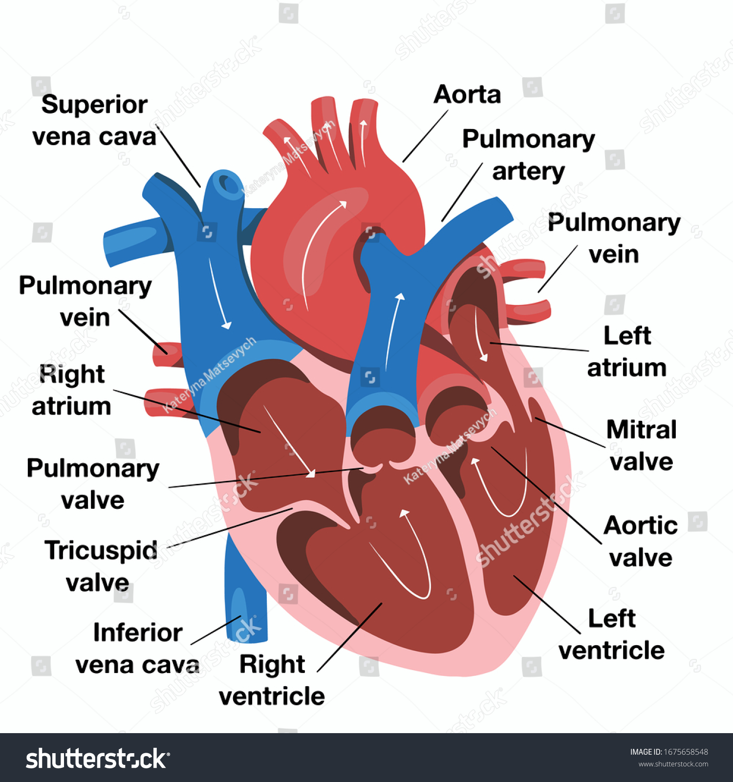
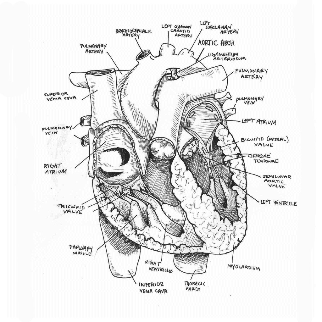
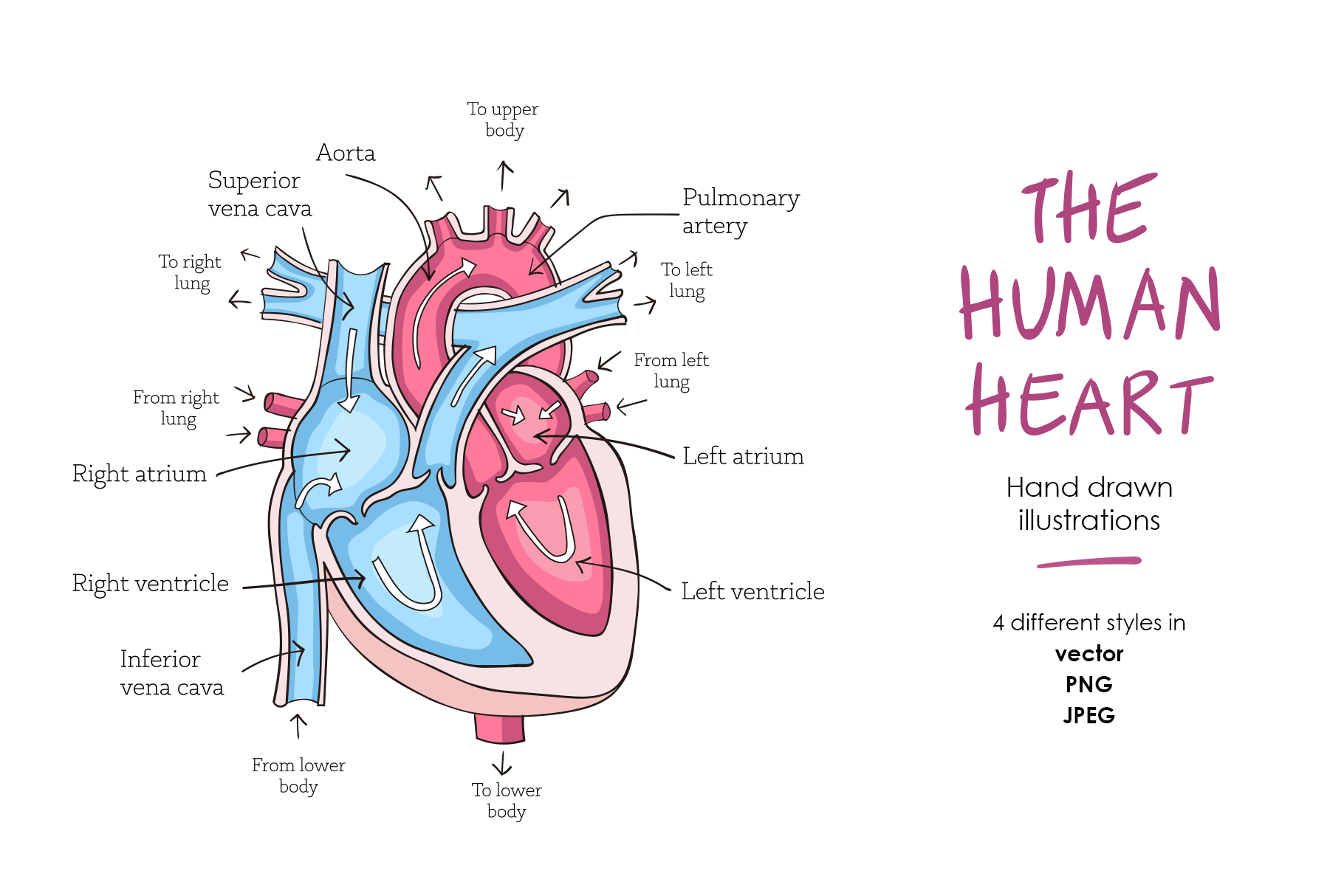
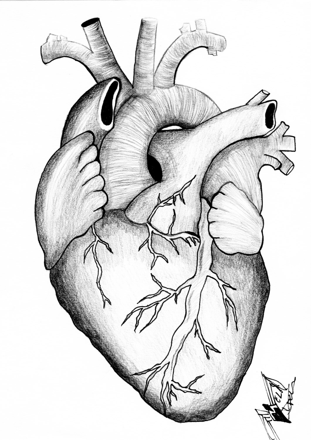
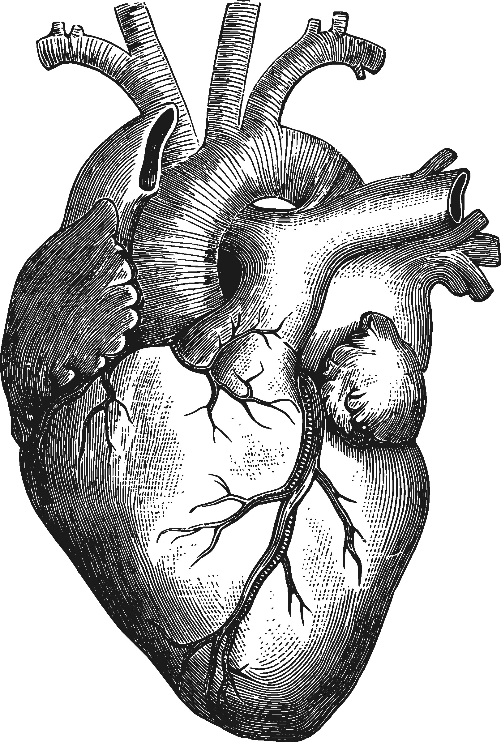
Anatomy Heart Drawing - Plus, you may just learn something new along the way. For this first step of our guide on how to draw a human heart, we will start with some outlines for the heart. Sketch out a basic outline of the heart, using our tutorial as a guide. Web drawing from the literature, the nominators assert that intraservice times are overvalued for these services and propose that these times should be adjusted to align more closely with average and/or typical surgery times. The user can show or hide the anatomical labels which provide a useful tool to create illustrations perfectly adapted for teaching. By following the simple steps, you too can easily draw a perfect human heart. Web best way to draw and label the heart! On its superior end, the base of the heart is attached to the aorta, pulmonary arteries and veins, and the vena cava. The outermost layer protects the heart and reduces friction with surrounding structures. Drawing a human heart is easier than you may think. I made this cool drawing as. Web these anatomical heart medical illustrations are highly detailed drawings that blend art with science. Web best way to draw and label the heart! We will then proceed to shape the heart, slowly refining it with our pencils into a. 20k views 4 years ago body part drawings: On its superior end, the base of the heart is attached to the aorta, pulmonary arteries and veins, and the vena cava. Web the intricate anatomy of the heart can be challenging to grasp, and so i hope you find this tool to be helpful in visualizing the cardiac system. How to draw women anatomy. Web function and anatomy of the heart made easy using labeled diagrams of cardiac structures and blood flow through the atria, ventricles, valves, aorta, pulmonary arteries veins, superior inferior vena cava, and chambers. For this first step of our guide on how to draw a human heart, we will start with some outlines for the heart. How to draw body parts. By following the simple steps, you too can easily draw a perfect human heart. Web these anatomical heart medical illustrations are highly detailed drawings that blend art with science. The videos and images on the atlas of human cardiac anatomy are free to download and use for educational purposes. The user can show or hide the anatomical labels which provide a useful tool to create illustrations perfectly adapted for teaching. Web the heart has three layers of tissue: Web best way to draw and label the heart! Web to draw the internal structure of the heart, start by sketching the 2 pulmonary veins to the lower left of the aorta and the bottom of the inferior vena cava slightly to the right of that. On its superior end, the base of the heart is attached to the aorta, pulmonary arteries and veins, and the vena cava. Web the heart is located in the thoracic cavity medial to the lungs and posterior to the sternum. Body reference poses plus size. Then, fill in the base of the heart with the right and left ventricles and the right and left atriums. The outermost layer protects the heart and reduces friction with surrounding structures. Use some curved lines for this aorta with a. Web best way to draw and label the heart! Drawing a human heart is easier than you may think. Body reference poses plus size. Web the intricate anatomy of the heart can be challenging to grasp, and so i hope you find this tool to be helpful in visualizing the cardiac system. Web the heart has three layers of tissue: By following the simple steps, you too can easily. On its superior end, the base of the heart is attached to the aorta, pulmonary arteries and veins, and the vena cava. The inferior tip of the heart, known as the apex, rests just superior to the diaphragm. Web drawings of the surface anatomy of the normal heart, anterior and posterior, with english labels. Some shading quickly helps to. Web. Web your heart sure does work hard, but that doesn’t mean you have to work hard to draw it! Female hand on hip pose drawing. Web best way to draw and label the heart! How to draw body parts. The outermost layer protects the heart and reduces friction with surrounding structures. Web your heart sure does work hard, but that doesn’t mean you have to work hard to draw it! 20k views 4 years ago body part drawings: The inferior tip of the heart, known as the apex, rests just superior to the diaphragm. ``do ponte osteotomies enhance correction in adolescent idiopathic scoliosis? Web the heart is located in the thoracic. The inferior tip of the heart, known as the apex, rests just superior to the diaphragm. Use some curved lines for this aorta with a. With this easy human heart drawing ideas, you can learn how to draw a human heart easily. On its superior end, the base of the heart is attached to the aorta, pulmonary arteries and veins,. Web the heart has three layers of tissue: Plus, you may just learn something new along the way. Body reference poses plus size. How to draw body parts. The outermost layer protects the heart and reduces friction with surrounding structures. Web the heart is located in the thoracic cavity medial to the lungs and posterior to the sternum. Plus, you may just learn something new along the way. The outermost layer protects the heart and reduces friction with surrounding structures. Web the heart has three layers of tissue: Web do you find the anatomy of the heart confusing? The innermost layer, provides a smooth lining for chambers and valves. Web to draw an anatomical heart realistically, pay attention to the proportions and positioning of the different parts of the heart, as well as their texture and color. The middle layer, composed of muscle tissue that enables heart contractions. ``do ponte osteotomies enhance correction in adolescent idiopathic scoliosis? By. By following the simple steps, you too can easily draw a perfect human heart. 48k views 1 year ago cardiovascular system. Web this interactive atlas of human heart anatomy is based on medical illustrations and cadaver photography. How to draw body parts. Web to draw an anatomical heart realistically, pay attention to the proportions and positioning of the different parts. Web function and anatomy of the heart made easy using labeled diagrams of cardiac structures and blood flow through the atria, ventricles, valves, aorta, pulmonary arteries veins, superior inferior vena cava, and chambers. Some shading quickly helps to. How to draw body parts. Included below are a magnificent color heart illustration, along with four monotype prints, which are possibly woodcuts, engravings, or lithographs. Web your heart sure does work hard, but that doesn’t mean you have to work hard to draw it! Web these anatomical heart medical illustrations are highly detailed drawings that blend art with science. 20k views 4 years ago body part drawings: Female hand on hip pose drawing. Web this interactive atlas of human heart anatomy is based on medical illustrations and cadaver photography. Web the intricate anatomy of the heart can be challenging to grasp, and so i hope you find this tool to be helpful in visualizing the cardiac system. The innermost layer, provides a smooth lining for chambers and valves. The videos and images on the atlas of human cardiac anatomy are free to download and use for educational purposes. Drawing a human heart is easier than you may think. Web learn how to draw a human heart with these 15 easy human heart drawing ideas with step by step sketch outline, printables and coloring pages. The outermost layer protects the heart and reduces friction with surrounding structures. I made this cool drawing as.Hand Drawn Illustration Human Heart Anatomy Stock Vector (Royalty Free
Sketch of human heart anatomy line and color on a checkered background
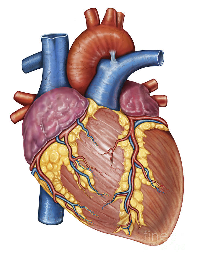
Gross Anatomy Of The Human Heart Digital Art by Stocktrek Images Pixels
Anatomical Drawing Heart at GetDrawings Free download
How to Draw the Internal Structure of the Heart 13 Steps
How to Draw the Internal Structure of the Heart (with Pictures)
Human heart anatomy (274491) Illustrations Design Bundles
Anatomical Heart Drawing at GetDrawings Free download
Anatomy of the human heart
Human Heart Drawing Images at Explore collection
Plus, You May Just Learn Something New Along The Way.
Web Best Way To Draw And Label The Heart!
Web The Heart Has Three Layers Of Tissue:
The Middle Layer, Composed Of Muscle Tissue That Enables Heart Contractions.
Related Post:
