Draw Heart Anatomy
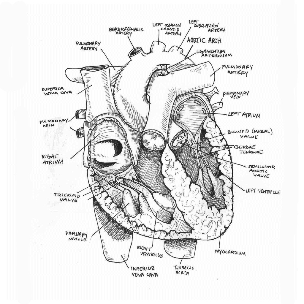
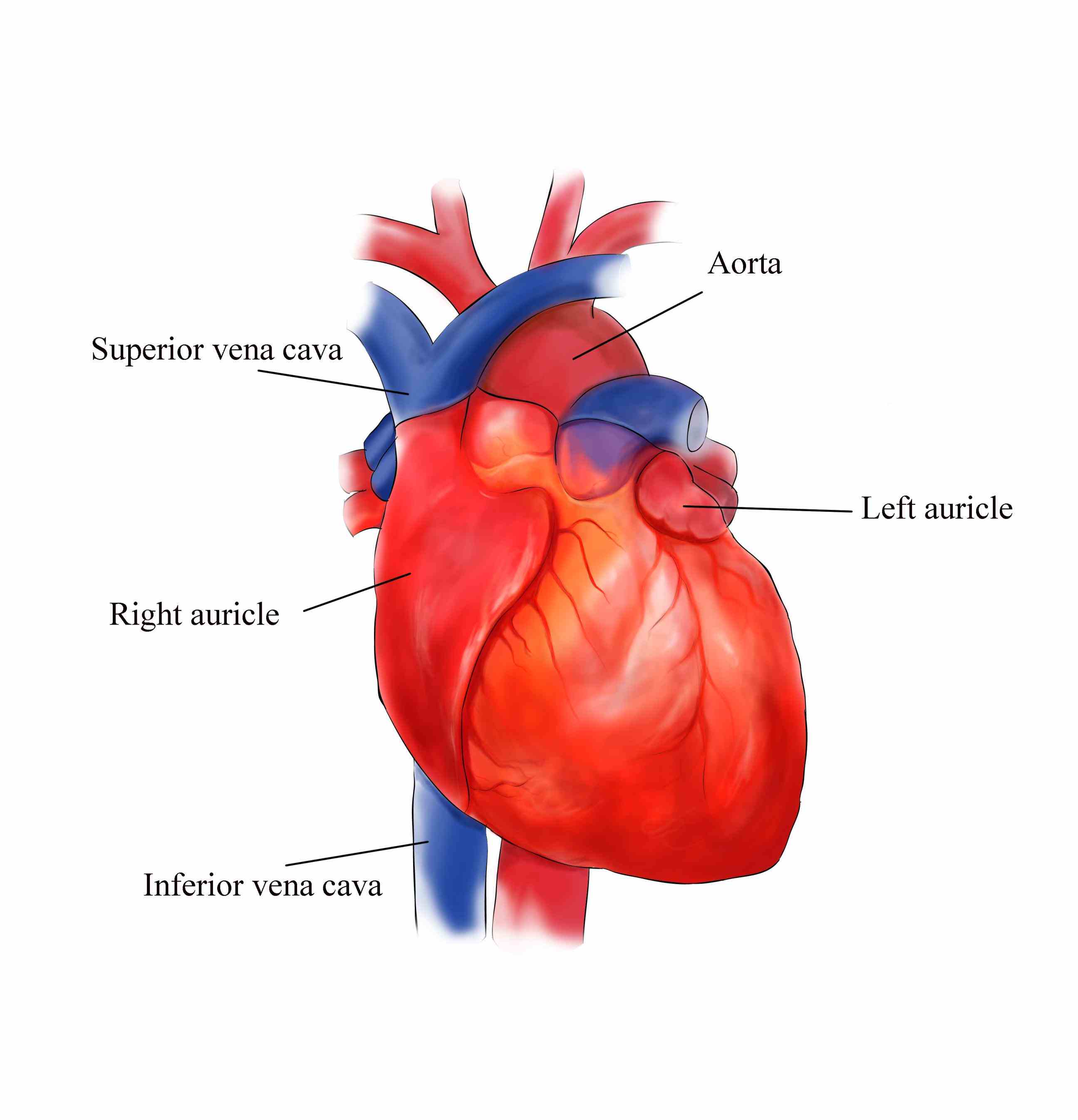
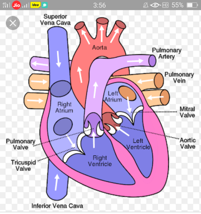
Draw Heart Anatomy - Sketch out a basic outline of the heart, using our tutorial as a guide. Web this interactive atlas of human heart anatomy is based on medical illustrations and cadaver photography. This key circulatory system structure is comprised of four chambers. In this lecture, dr mike shows the two best ways to draw and. Web dr matt & dr mike. The valves of the heart. The inferior tip of the heart, known as the apex, rests just superior to the diaphragm. Some shading quickly helps to. Web the heart is located in the thoracic cavity medial to the lungs and posterior to the sternum. Web to draw an anatomical heart realistically, pay attention to the proportions and positioning of the different parts of the heart, as well as their texture and color. Web heart, organ that serves as a pump to circulate the blood. The valves of the heart. What is the function of the heart? It also takes away carbon dioxide and other waste so other organs can dispose of them. This key circulatory system structure is comprised of four chambers. We will then proceed to shape the heart, slowly refining it with our pencils into a. What does the heart look like and how does it work? Web your heart sure does work hard, but that doesn’t mean you have to work hard to draw it! Rotate the 3d model to see how the heart's valves control blood flow between heart chambers and blood flow out of the heart. Plus, you may just learn something new along the way. Drag and drop the text labels onto the boxes next to the heart diagram. In this lecture, dr mike shows the two best ways to draw and. Web anatomy of the interior of the heart. Your heart’s main function is to move blood throughout your body. Cardiovascular system animation for u. Medical conditions such as heart conditions or intellectual and developmental disabilities are common. Web heart, organ that serves as a pump to circulate the blood. This key circulatory system structure is comprised of four chambers. It consists of four main chambers: On its superior end, the base of the heart is attached to the aorta, pulmonary arteries and veins, and the vena cava. 48k views 1 year ago cardiovascular system. Web the heart is a muscular organ that pumps blood around the body by circulating it through the circulatory/vascular system. Blood brings oxygen and nutrients to your cells. Two atria and two ventricles. Rotate the 3d model to see how the heart's valves control blood flow between heart chambers and blood flow out. In this interactive, you can label parts of the human heart. Your heart’s main function is to move blood throughout your body. It consists of four main chambers: Medical conditions such as heart conditions or intellectual and developmental disabilities are common. This image shows the four chambers of the heart and the direction that blood flows through the heart. Web the heart is a muscular organ that pumps blood around the body by circulating it through the circulatory/vascular system. The inferior tip of the heart, known as the apex, rests just superior to the diaphragm. Web the heart is a mostly hollow, muscular organ composed of cardiac muscles and connective tissue that acts as a pump to distribute blood. The valves of the heart. Drag and drop the text labels onto the boxes next to the heart diagram. What does the heart look like and how does it work? Rotate the 3d model to see how the heart's valves control blood flow between heart chambers and blood flow out of the heart. The heart is a conical hollow muscular. Web the surfaces and borders of the heart. What does the heart look like and how does it work? In this interactive, you can label parts of the human heart. Web anatomy of the heart made easy along with the blood flow through the cardiac structures, valves, atria, and ventricles. Web heart, organ that serves as a pump to circulate. The heart is an amazing organ. Rotate the 3d model to see how the heart's valves control blood flow between heart chambers and blood flow out of the heart. This key circulatory system structure is comprised of four chambers. This image shows the four chambers of the heart and the direction that blood flows through the heart. The effects of. The user can show or hide the anatomical labels which provide a useful tool to create illustrations perfectly adapted for teaching. The heart is a conical hollow muscular organ situated in the middle mediastinum and is enclosed within the pericardium. Drag and drop the text labels onto the boxes next to the heart diagram. Cardiovascular system animation for u. Web. Web the heart is a mostly hollow, muscular organ composed of cardiac muscles and connective tissue that acts as a pump to distribute blood throughout the body’s tissues. Controls the rhythm and speed of your heart rate. Web the heart is located in the thoracic cavity medial to the lungs and posterior to the sternum. This image shows the four. Web to draw the internal structure of the heart, start by sketching the 2 pulmonary veins to the lower left of the aorta and the bottom of the inferior vena cava slightly to the right of that. Web function and anatomy of the heart made easy using labeled diagrams of cardiac structures and blood flow through the atria, ventricles, valves,. Web to draw an anatomical heart realistically, pay attention to the proportions and positioning of the different parts of the heart, as well as their texture and color. Understanding its basic anatomy is crucial to understanding how it functions. This website is aimed to be an easily accessible web based app that features an on demand high fidelity rendering of. The user can show or hide the anatomical labels which provide a useful tool to create illustrations perfectly adapted for teaching. Medical conditions such as heart conditions or intellectual and developmental disabilities are common. Blood brings oxygen and nutrients to your cells. Web your heart sure does work hard, but that doesn’t mean you have to work hard to draw it! Plus, you may just learn something new along the way. What does the heart look like and how does it work? Drag and drop the text labels onto the boxes next to the heart diagram. Web the surfaces and borders of the heart. Rotate the 3d model to see how the heart's valves control blood flow between heart chambers and blood flow out of the heart. Your heart’s main function is to move blood throughout your body. Monolids and other physical characteristics often occur with down syndrome, a genetic disorder also called trisomy 21. Anatomical areas the anatomical snuffbox. Web dr matt & dr mike. In this interactive, you can label parts of the human heart. Web function and anatomy of the heart made easy using labeled diagrams of cardiac structures and blood flow through the atria, ventricles, valves, aorta, pulmonary arteries veins, superior inferior vena cava, and chambers. It consists of four main chambers:Anatomical Drawing Heart at GetDrawings Free download
How to Draw the Internal Structure of the Heart (with Pictures)
How to draw Human Heart with colour Human Heart labelled diagram
How to Draw Human Heart Diagram Drawing / easy way Step by step YouTube
Sketch of human heart anatomy ,line and color on a checkered background
External Structure Of Heart Anatomy Diagram
Heart Anatomy · Anatomy and Physiology
How To Draw Human Heart Diagram
How to Draw the Internal Structure of the Heart 13 Steps
anatomically correct human heart by NIku Arbabi embroidery
This Website Is Aimed To Be An Easily Accessible Web Based App That Features An On Demand High Fidelity Rendering Of The Human Heart To Use While On Rounds, In Teaching Conferences, Or By Bedside.
Web This Interactive Atlas Of Human Heart Anatomy Is Based On Medical Illustrations And Cadaver Photography.
The Effects Of Alcohol Exposure On The Developing Fetus Can Include A Wide.
Then, Fill In The Base Of The Heart With The Right And Left Ventricles And The Right And Left Atriums.
Related Post:


