Draw The F As Seen In The Low Power Field
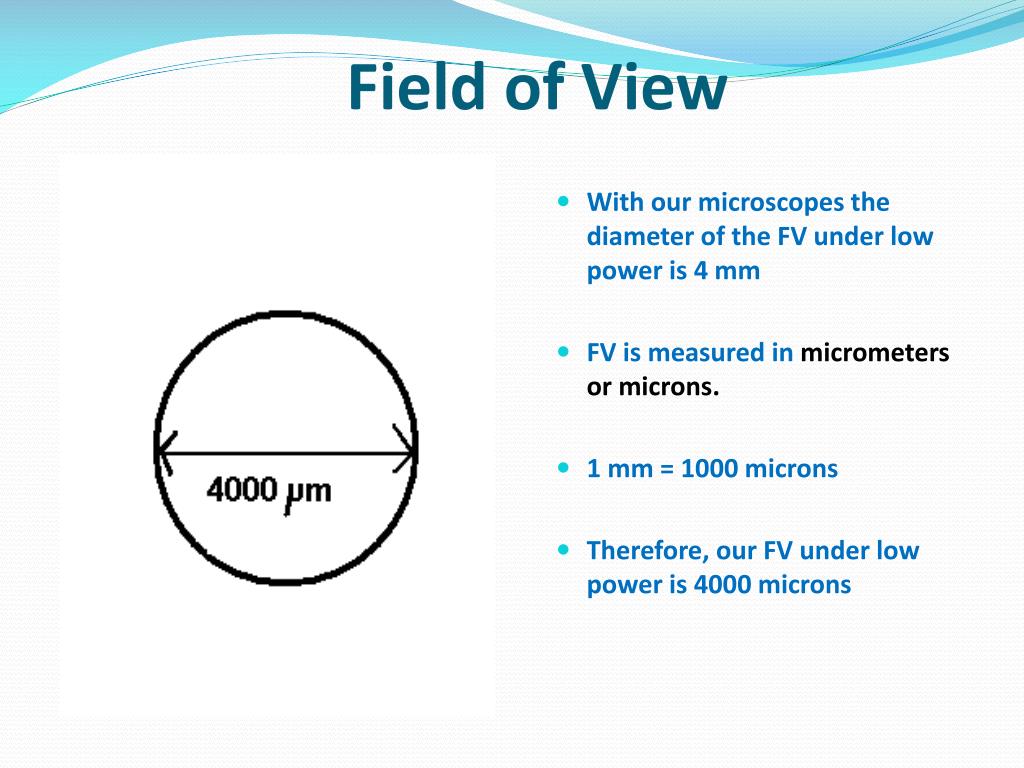
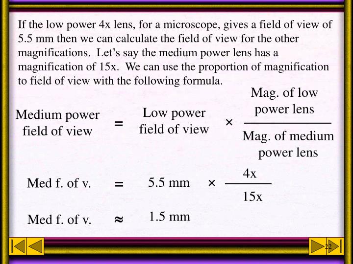
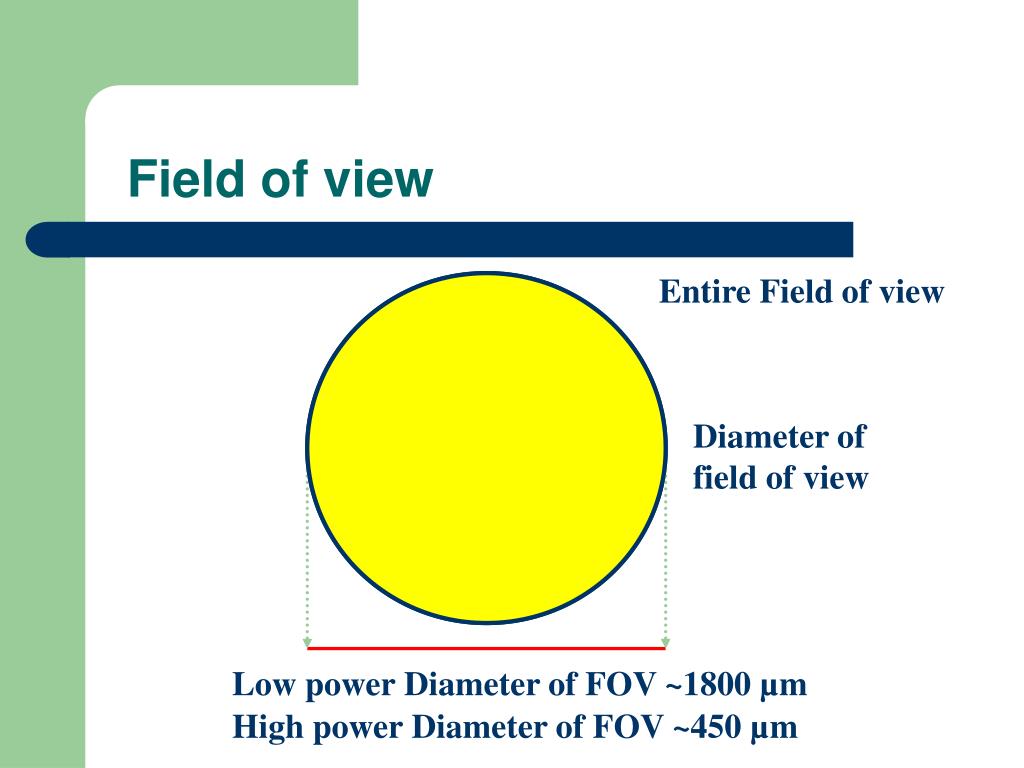
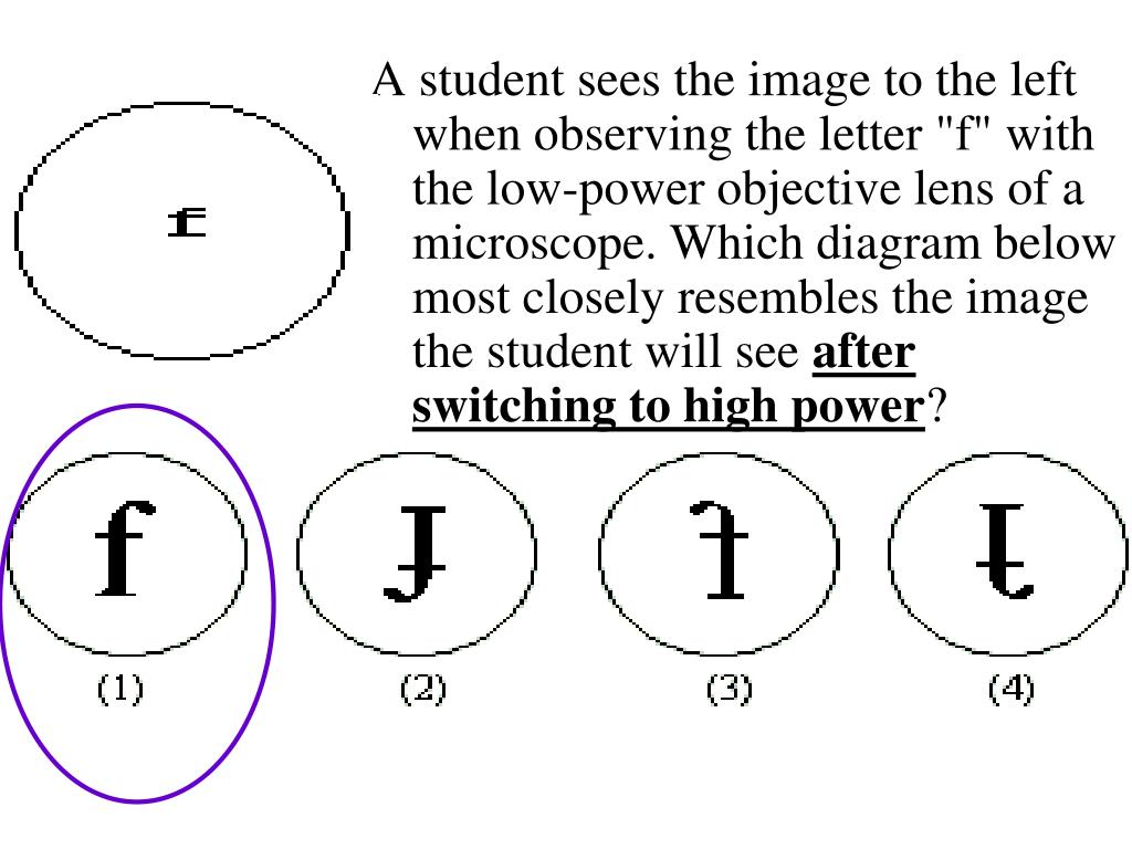
Draw The F As Seen In The Low Power Field - Web study with quizlet and memorize flashcards containing terms like define total magnification, explain the proper technique for transporting the microscope, true or. Carefully draw your elodea at all three magnifications. Used color pencils to show how the stain appears. Calculate the total magnification of the low. Web the fields of view and approximate distances across for scanning, low, and high power are as follows: You have been asked to. Web in the circle below. You have been asked to prepare a slide with the letter $f$ on it (as. You have been a ked to prepare slide with the letter. Copy the following 2 tables into your notes under the “observations” section of your lab report. Place a transparent metric ruler under the low power (lp) objective of a microscope. Your cheek cells should look like pancake shapes with uneven edges. If you cannot see anything, move the slide slightly while viewing and focusing. It may appear darker or lighter in spots. Web you have been asked to prepare a slide with the letter f on it (as shown below). Your solution’s ready to go!. Carefully draw your elodea at all three magnifications. In order to measure the cells under the. Total magnification = field diameter = 1.6 mm. You have been asked to. Estimate the length (longest dimension) of the object in urn: In order to measure the cells under the. If you cannot see anything, move the slide slightly while viewing and focusing. Use shading to show darker and. Web in the circle below. Your cheek cells should look like pancake shapes with uneven edges. You have been a ked to prepare slide with the letter. Use the focusing sequence to view the slide under low power. Obtain a microscope and bring it to the laboratory bench. Web field of view (fov) in a microscope, we ordinarily observe things within a circular space (or field) as defined by the lenses. Estimate the length (longest dimension) of the object in $\mu \mathrm. Place a transparent metric ruler under the low power (lp) objective of a microscope. It may appear darker or lighter in spots. Estimate the number of millimeters that. Your solution’s ready to go!. Carefully draw your elodea at all three magnifications. In order to measure the cells under the. You have been asked to. It may appear darker or lighter in spots. Draw the letter “e” as it. Web in the circle below. Calculate the total magnification of the low. Estimate the number of millimeters that. If you cannot see anything, move the slide slightly while viewing and focusing. Total magnification = field diameter = 1.6 mm. Use shading to show darker and. Calculate the total magnification of the low. We refer to this observable area as the field of view. Obtain a microscope and bring it to the laboratory bench. Estimate the length (longest dimension) of the object in $\mu \mathrm. Estimate the length (longest dimension) of the object in urn: Total magnification = field diameter = 1.6 mm. Web on low power only, use the coarse focus knob to get the object into focus. You have been asked to. Copy the following 2 tables into your notes under the “observations” section of your lab report. Use shading to show darker and. Web leaving the ruler in place, rotate the low power (10x) objective into position. Don’t forget to label the tables! Estimate the length (longest dimension) of the object in urn: Web _________________________________ what are the numerous small, green structures? Your solution’s ready to go!. Your cheek cells should look like pancake shapes with uneven edges. You have been a ked to prepare slide with the letter. Place a transparent metric ruler under the low power (lp) objective of a microscope. Total magnification = field diameter = 1.6 mm. We refer to this observable area as the field of view. In order to measure the cells under the. Web draw your specimen as it appears under low power. You have been asked to. It may appear darker or lighter in spots. We refer to this observable area as the field of view. Draw the letter “e” as it. You have been a ked to prepare slide with the letter. Web study with quizlet and memorize flashcards containing terms like define total magnification, explain the proper technique for transporting the microscope, true or. Carefully draw your elodea at all three magnifications. Use the focusing sequence to view the slide under low power. Web draw your specimen as it appears under low power. Web view the slide with your eyes, and then place it onto the microscope. Web in the circle below. Web study with quizlet and memorize flashcards containing terms like define total magnification, explain the proper technique for transporting the. Web in the circle below. If you cannot see anything, move the slide slightly while viewing and focusing. (use the proper transport technique!) compare your microscope with the figure on the following page and identify. Obtain a microscope and bring it to the laboratory bench. Web on low power only, use the coarse focus knob to get the object into focus. Ned mated diameter of mm 7. Carefully draw your elodea at all three magnifications. You have been a ked to prepare slide with the letter. Estimate the length (longest dimension) of the object in urn: Copy the following 2 tables into your notes under the “observations” section of your lab report. You have been asked to. In order to measure the cells under the. Your cheek cells should look like pancake shapes with uneven edges. Total magnification = field diameter = 1.6 mm. It may appear darker or lighter in spots. Use shading to show darker and.Figure 10 from LowPower FieldProgrammable VLSI Using Multiple Supply
PPT The Microscope PowerPoint Presentation, free download ID6006599
PPT Living Organisms PowerPoint Presentation ID238651
PPT Introduction to the Light Microscope PowerPoint Presentation
Pathological findings. Lowpower field (a) and highpower field (b) of
Low power plan diagram AS Level Biology YouTube
PPT Microscope Review PowerPoint Presentation, free download ID6690365
PPT Microscope Review PowerPoint Presentation, free download ID6690365
Figure2.It shows macro specimen (A), lowpower field (B), and
How to design progressive lenses Knowledgebase
Calculate The Total Magnification Of The Low.
Use The Focusing Sequence To View The Slide Under Low Power.
Use The Fine Focus If Necessary To Bring The Ruler Into Focus.
Web You Have Been Asked To Prepare A Slide With The Letter F On It (As Shown Below).
Related Post:


