Drawing Of Skeletal Muscle
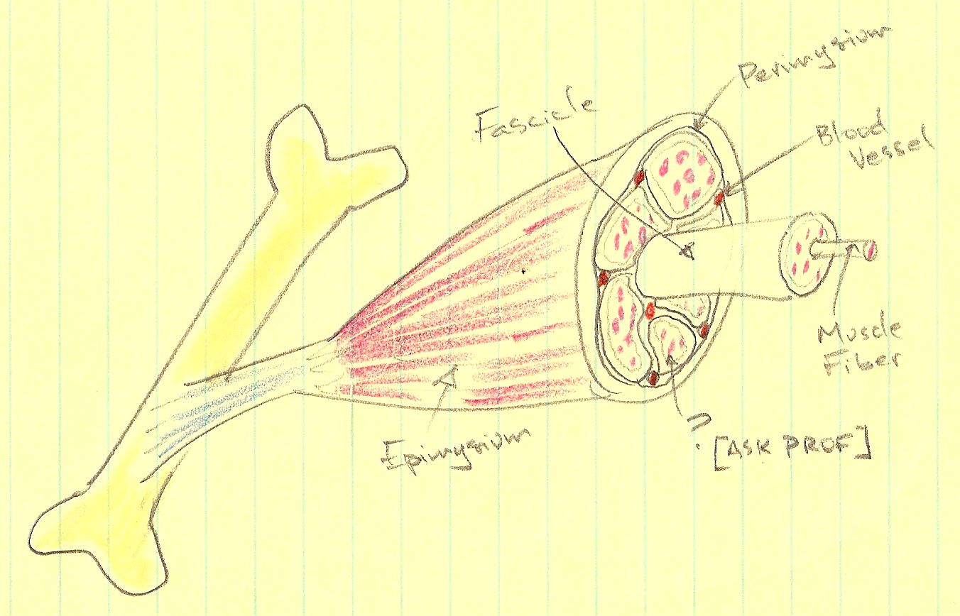
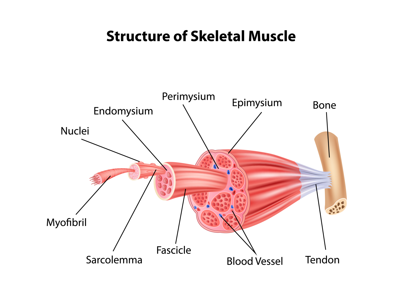
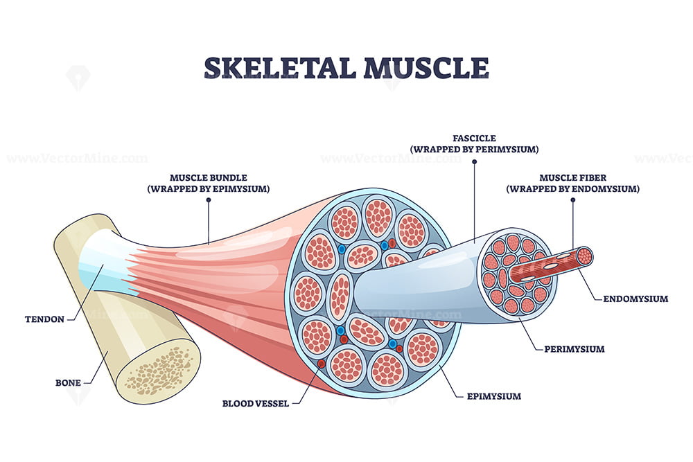
Drawing Of Skeletal Muscle - Understanding the anatomy of muscles is essential for realistic depictions. They are made of muscle fibres and play an important role in muscle excitation and contraction. Within muscles, there are layers of connective tissue called the epimysium, perimysium, and endomysium. Explain how muscles work with tendons to move the body. We’ve created muscle anatomy charts for every muscle containing region of the body: There are more than 600 skeletal muscles, and they makes up about 40 percent of a person’s body weight. Each chart groups the muscles of that region into its component groups, making your revision a million times easier. Web #muscle #diagram #howtodrawthis is a drawing of structure of the skeletal muscle. The bones of the skeletal system serve to protect the body's organs, support the weight of the body, and give the body shape. Web skeletal muscle is an excitable, contractile tissue responsible for maintaining posture and moving the orbits, together with the appendicular and axial skeletons. There are three types of muscles: Web skeletal muscle is a specialized contractile tissue found in animals which functions to move an organism’s body. Tendons (tough bands of connective tissue) attach skeletal muscle tissue to bones throughout your body. A comprehensive guide to drawing realistic muscles. These videos will help you to draw these diagram. The bones of the skeletal system serve to protect the body's organs, support the weight of the body, and give the body shape. Understanding the anatomy of muscles is essential for realistic depictions. Blood vessels and nerves enter the connective tissue and branch in the cell. They make up between 30 to 40% of your total body mass. Muscles work on a macro level, starting with tendons that attach muscles to bones. Web in the musculoskeletal system, the muscular and skeletal systems work together to support and move the body. They make up between 30 to 40% of your total body mass. Web skeletal muscles contain connective tissue, blood vessels, and nerves. Each of these muscles is a discrete organ constructed of skeletal muscle tissue, blood vessels, tendons, and nerves. A comprehensive guide to drawing realistic muscles. Web knowing these skeletal basics is essential before delving into muscles. The fibers run the entire length of the muscle they come from and so are usually too long to have their ends visible when viewed under the microscope. Each chart groups the muscles of that region into its component groups, making your revision a million times easier. It is the most common muscle tissue. You can draw the structure of the muscle for your exams and projects by us. They are made of muscle fibres and play an important role in muscle excitation and contraction. A comprehensive guide to drawing realistic muscles. The bones of the skeletal system serve to protect the body's organs, support the weight of the body, and give the body shape. There are three layers of connective tissue: It is the most common muscle tissue. Web different type of muscle cells have different unique characteristics. These layers cover muscle subunits, individual muscle cells, and myofibrils respectively. Web skeletal muscles are voluntary and striated in nature. For example, the skeletal muscle is the only type of muscle cell that is always multinucleated (for more info see the latter half of sal's video). It is the most. Each of these muscles is a discrete organ constructed of skeletal muscle tissue, blood vessels, tendons, and nerves. Web #muscle #diagram #howtodrawthis is a drawing of structure of the skeletal muscle. Web skeletal muscles are voluntary and striated in nature. Web drawing from the literature, the nominators assert that intraservice times are overvalued for these services and propose that these. Web #muscle #diagram #howtodrawthis is a drawing of structure of the skeletal muscle. Web skeletal muscle is a specialized contractile tissue found in animals which functions to move an organism’s body. Web these tissues include the skeletal muscle fibers, blood vessels, nerve fibers, and connective tissue. Web each skeletal muscle has three layers of connective tissue (called “mysia”) that enclose. Web hello friends, this is my youtube channel and in this channel i used to share videos of different diagrams in easy way and step by step tutorials. We’ve created muscle anatomy charts for every muscle containing region of the body: Web skeletal muscles are voluntary and striated in nature. Web a helpful and detailed muscle manual by artist eridey.. You can draw the structure of the muscle for your exams and projects by us. There are three types of muscles: The majority of the muscles in your body are skeletal muscles. Skeletal muscle fibers are organized into groups called fascicles. Identify areas of the skeletal muscle fibers. There are three types of muscles: It attaches to bones and the orbits through tendons. You can draw the structure of the muscle for your exams and projects by us. Skeletal muscle fibers are organized into groups called fascicles. Web a helpful and detailed muscle manual by artist eridey. It consists of long, parallel multinucleate cells bundled together by collagenous sheaths and through this regular organization allow the skeletal muscles to generate powerful contractions, along with a power output up to 100 watts per kilogram of tissue. Web a complete list of muscles. Web different type of muscle cells have different unique characteristics. After all, they offer anchor points. Skeletal muscle fibers are organized into groups called fascicles. Web skeletal muscles are striated and voluntary. Web in this video i have shown the simplest way of drawing muscle drawing. Web a helpful and detailed muscle manual by artist eridey. Skeletal muscle is comprised from a series of bundles of muscle fibers, surrounded by protective membranes. They are made of muscle fibres and play an important role in muscle excitation and contraction. The majority of the muscles in your body are skeletal muscles. A comprehensive guide to drawing realistic muscles. Identify areas of the skeletal muscle fibers. Blood vessels and nerves enter the connective tissue and branch in the cell. Blood vessels and nerves enter the connective tissue and branch in the cell. Within muscles, there are layers of connective tissue called the epimysium, perimysium, and endomysium. Web skeletal muscle is a specialized contractile tissue found in animals which functions to move an organism’s body. You can draw the structure of the muscle for your exams and projects by us. Web #muscle #diagram #howtodrawthis is a drawing of structure of the skeletal muscle. It is the pen diagram of skeletal, smooth and cardiac muscle for class 10, 11 and 12. These videos will help you to draw these diagram. Web skeletal muscles contain connective tissue, blood vessels, and nerves. Web each skeletal muscle has three layers of connective tissue (called “mysia”) that enclose it and provide structure to the muscle as a whole, and also compartmentalize the muscle fibers within the muscle (figure 10.3). Understanding the anatomy of muscles is essential for realistic depictions. Web drawing from the literature, the nominators assert that intraservice times are overvalued for these services and propose that these times should be adjusted to align more closely with average and/or typical surgery times. The majority of the muscles in your body are skeletal muscles. Web practice drawing different muscle groups, including the biceps, triceps, abs, and quads. Identify areas of the skeletal muscle fibers. Web in this video i have shown the simplest way of drawing muscle drawing. Describe the layers of connective tissues packaging skeletal muscle.Illustration of Structure Skeletal Muscle Anatomy Stock Photo Alamy
Muscle Tissue Drawing at GetDrawings Free download
How To Draw Skeletal, Smooth and Cardiac Muscle Diagram Types Of
Skeletal Muscle Tissue Labeled Sarcolemma
Skeletal Muscle Drawing at Explore collection of
How To Draw Structure Of Skeletal Muscle YouTube in 2022 Biology
Structure Skeletal Muscle Anatomy by Tigatelu on Dribbble
Skeletal muscle structure with anatomical inner layers outline diagram
Skeletal muscle description with cross section structure outline
Skeletal Muscle Diagram 101 Diagrams
Web A Complete List Of Muscles.
Web In The Musculoskeletal System, The Muscular And Skeletal Systems Work Together To Support And Move The Body.
For Example, The Skeletal Muscle Is The Only Type Of Muscle Cell That Is Always Multinucleated (For More Info See The Latter Half Of Sal's Video).
They Are Made Of Muscle Fibres And Play An Important Role In Muscle Excitation And Contraction.
Related Post:


