Drawing Of Tissue
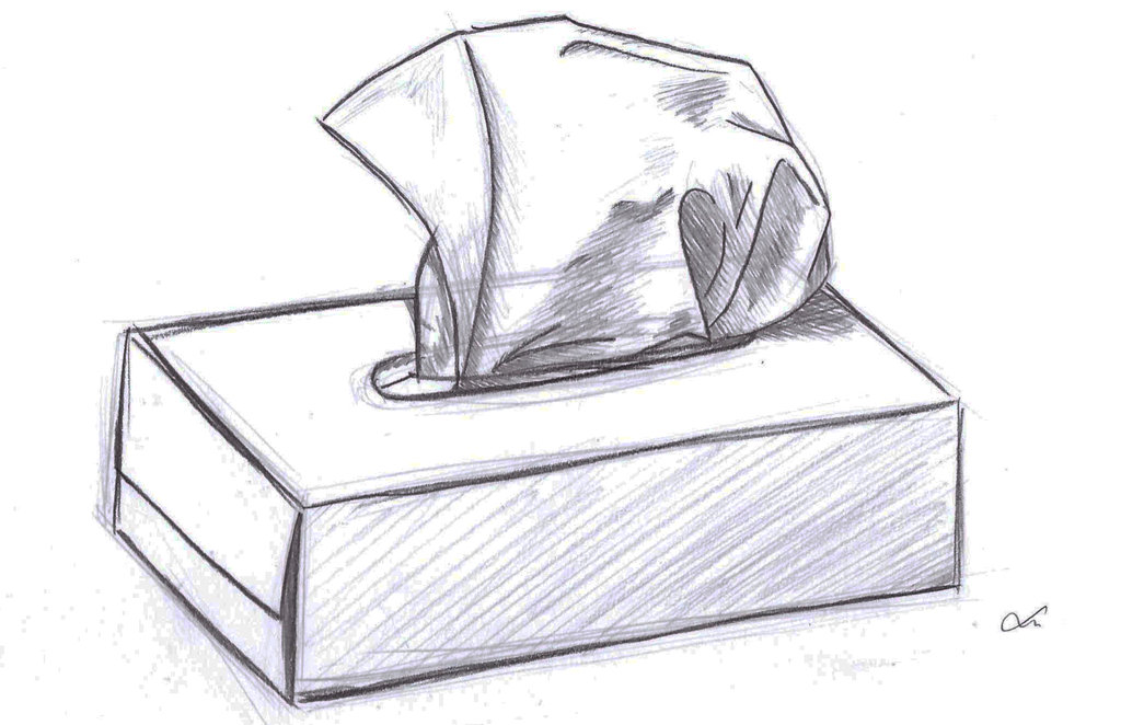
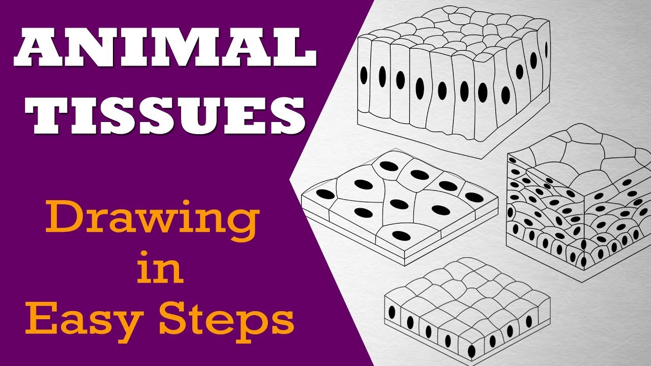
Drawing Of Tissue - Microscopic observation reveals that the cells in a tissue share morphological features and are arranged in an orderly pattern that achieves the tissue’s functions. Web epithelial tissue serves two main functions in the body. Draw leaves on the folded edge. Web the term tissue is used to describe a group of cells found together in the body. The standard tools for studying tissues is by embedding and sectioning using the paraffin block. Web are you looking for the best images of tissue drawing? Connective, muscle, nervous, and epithelial. Web tissues are groups of cells that have a similar structure and act together to perform a specific function. I strongly suggest using a companion atlas or digital resource as you work through these exercises. It provides linings for external and internal surfaces that face harsh environments. The primary tissue types work together to contribute to the overall. Gather your supplies and materials. These categories are epithelial, connective, muscle, and nervous. The standard tools for studying tissues is by embedding and sectioning using the paraffin block. Epithelial tissue, connective tissue, muscle tissue, and nervous tissue. There are no labeled images here, as the goal is to get you to practice identifying structures on your own. The outer layer of the skin is epithelial tissue, as are the innermost layers of the digestive tract, the respiratory tract, and blood vessels. Web epithelial tissue serves two main functions in the body. The cells within a tissue share a common embryonic origin. This is made up of thin, flat and hexagonal cells. This facility is the largest publicly held plant. Drawings can highlight the important features of a specimen. The standard tools for studying tissues is by embedding and sectioning using the paraffin block. Web let us learn in detail about the types of tissues in different organs. Epithelial tissue, connective tissue, muscle tissue, and nervous tissue. Web this female anatomy diagram is a good place to start if you're unsure of exactly where parts of the female reproductive and urinary systems are in comparison to one another. Web choose from drawing of tissue stock illustrations from istock. Web come join and follow us to learn how to draw. Web how to draw different types of epithelial tissue | animal tissue drawing | craft magician.drawn by: There are no labeled images here, as the goal is to get you to practice identifying structures on your own. Drawings can highlight the important features of a specimen. The following sections go into detail about these and. Web the term tissue is used to describe a group of cells found together in the body. Web are you looking for the best images of tissue drawing? The standard tools for studying tissues is by embedding and sectioning using the paraffin. Web simple squamous epithelium: I strongly suggest using a companion atlas or digital resource as you work through these exercises. Web the term tissue is used to describe a group of cells found together in the body. The cells within a tissue share a common embryonic origin. Web tissues are groups of cells that have a similar structure and act. Epithelial tissue, connective tissue, muscle tissue, and nervous tissue. Connective, muscle, nervous, and epithelial. Web these lab worksheets are designed to guide you through a laboratory experience in histology. Possible uses include drawing clear skies, skin tones, or luminous flowers. I strongly suggest using a companion atlas or digital resource as you work through these exercises. Drawing is a very important skill in biology and is considered a type of data collection because drawings help to record data from specimens. The cells within a tissue share a common embryonic origin. Web this female anatomy diagram is a good place to start if you're unsure of exactly where parts of the female reproductive and urinary systems are. This facility is the largest publicly held plant. Animal tissues in easy steps and compact way. Web choose from tissue drawing stock illustrations from istock. The cells within a tissue share a common embryonic origin. Web there are four basic tissue types defined by their morphology and function: Fahmida islam moon.this video helps you to draw science. These categories are epithelial, connective, muscle, and nervous. You can also cut longer strips of tissue paper, depending on what look you're going for. I strongly suggest using a companion atlas or digital resource as you work through these exercises. The standard tools for studying tissues is by embedding and sectioning. Drawing is a very important skill in biology and is considered a type of data collection because drawings help to record data from specimens. Web drawings of cells are typically made when visualizing cells at a higher magnification power, whereas plan drawings are typically made of tissues viewed under lower magnifications (individual cells are never drawn in a plan diagram). Simply subscribe us for more drawing tutorial. I also suggest keeping a notebook (either. Web this female anatomy diagram is a good place to start if you're unsure of exactly where parts of the female reproductive and urinary systems are in comparison to one another. Cut the bleeding tissue paper into small squares. Web are you looking for the best. Possible uses include drawing clear skies, skin tones, or luminous flowers. It provides linings for external and internal surfaces that face harsh environments. Web simple squamous epithelium: Microscopic observation reveals that the cells in a tissue share morphological features and are arranged in an orderly pattern that achieves the tissue’s functions. Cut the bleeding tissue paper into small squares. The following sections go into detail about these and. There are no labeled images here, as the goal is to get you to practice identifying structures on your own. Microscopic observation reveals that the cells in a tissue share morphological features and are arranged in an orderly pattern that achieves the tissue’s functions. Web are you looking for the best. Drawings can highlight the important features of a specimen. The cells within a tissue share a common embryonic origin. I strongly suggest using a companion atlas or digital resource as you work through these exercises. Possible uses include drawing clear skies, skin tones, or luminous flowers. Web tissues are groups of cells that have a similar structure and act together to perform a specific function. There are four different types of tissues in animals: Web connective tissue has the most types of subcategories and the most varied functions of all the four major tissue types (epithelial, muscular, nervous, and connective tissues.) bone and cartilage are connective tissues, as. The following sections go into detail about these and. Web epithelial tissue serves two main functions in the body. These categories are epithelial, connective, muscle, and nervous. Draw leaves on the folded edge. Epithelial tissue creates protective boundaries and is involved in the diffusion of ions and molecules. Connective, muscle, nervous, and epithelial. Web the term tissue is used to describe a group of cells found together in the body. Web this female anatomy diagram is a good place to start if you're unsure of exactly where parts of the female reproductive and urinary systems are in comparison to one another. A large central rounded nucleus.How to Draw Tissue box Step by Step YouTube
Tissue Drawing at Explore collection of Tissue Drawing
How to Draw a Tissue Box 6 Steps (with Pictures) wikiHow
Reticular Connective Tissue Drawing Master the Art of Illustrating
Tissue Box Drawing at Explore collection of Tissue
Tissue Drawing at Explore collection of Tissue Drawing
drawing of a tissue box kaileighj
Transparent Body Tissues Clipart Squamous Epithelium Png Download
epithelial tissue, drawing Stock Image C015/2525 Science Photo
How to draw VARIOUS TYPES OF SIMPLE TISSUES class 9 science YouTube
Web Come Join And Follow Us To Learn How To Draw.
Drawing Is A Very Important Skill In Biology And Is Considered A Type Of Data Collection Because Drawings Help To Record Data From Specimens.
The Outer Layer Of The Skin Is Epithelial Tissue, As Are The Innermost Layers Of The Digestive Tract, The Respiratory Tract, And Blood Vessels.
Web Drawings Of Cells Are Typically Made When Visualizing Cells At A Higher Magnification Power, Whereas Plan Drawings Are Typically Made Of Tissues Viewed Under Lower Magnifications (Individual Cells Are Never Drawn In A Plan Diagram)
Related Post:


